Receptor location plays a key role in their function
A research team led by Prof. Martin Lohse has identified, for the first time, where special receptors are located on heart muscle cells. The findings open up new perspectives for developing therapies for chronic heart failure.

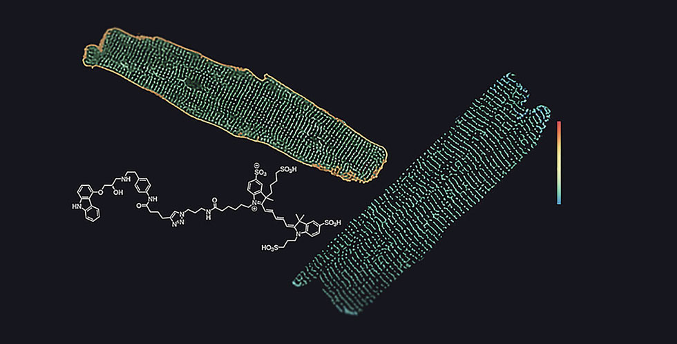
Beta1- and beta2-adrenergic receptors in heart muscle cells (cardiomyocytes): In the left cell, beta1-receptors are labelled – they can be found both at the cell surface (yellow) and in the internal t-tubules (green). In the right cell, the beta2-receptors are labelled– they occur only in the t-tubules (green), but not at the cell surface (which is, therefore, not visible). (Image: Marc Bathe-Peters & Horst-Holger Boltz)
In the heart there are two different subtypes of beta-adrenergic receptors -- beta1 and beta2 -- which are activated by the stress hormones adrenaline and noradrenaline. They both trigger the strongest stimulation of the heart rate and pumping capacity that we know of. The two subtypes are highly similar biochemically, but differ substantially in terms of their functional and therapeutic relevance.
Both receptor types can stimulate the heart in the short term, yet when the beta1 receptor is activated over a prolonged period of time, it has a range of effects that are not seen with beta2. Beta1 can elicit a number of persistent changes and is endowed with the ability to initiate -- often detrimental -- growth of the heart muscle cells by activating various genes.
Recent studies by researchers at the Max Delbrück Center for Molecular Medicine in the Helmholtz Association (MDC) in Berlin, the University of Erlangen, and ISAR Bioscience in Munich-Planegg have now shed light on the mechanisms behind these different effects. The research teams have published the results of their work in the current issue of the journal Proceedings of the National Academy of Sciences.
Special ligands and new microscopy methods
“Using a fluorescent ligand synthesized at the University of Erlangen and novel highly sensitive microscopy methods, we were able to show for the first time where these receptors are located on heart muscle cells,” explains Professor Martin Lohse, Chairman of ISAR Bioscience. “The endogenous receptors are expressed at relatively low levels,” explains Dr. Paolo Annibale, long-term collaborator of Prof. Lohse and co-lead author of the study. “To detect their movement, it was necessary to use a form of spectroscopy based on the analysis of the signal’s minute fluorescence fluctuations.”
This revealed that beta1 receptors are found on the entire surface of heart muscle cells, while beta2 receptors are exclusively found in specific structures in these cells called T-tubules. Through invaginations of the cell surface, these tubules create a pipe-like network that runs through the entire interior of heart muscle cells. “One of the research focuses of our team at the MDC is the relationship between receptor function and subcellular localization,” adds Annibale. “So the biophysical environment of T-tubules, which have curved membranes, is of particular interest to us.”
Not all heart muscle cells have beta1 receptors
“The specific cellular location of beta2 receptors explains why they have a much narrower range of functionality than beta1 receptors and why they are limited to direct and short-term stimulation of the heart,” explains Lohse. Such stimulation is mediated by signals that are locally restricted to the cell membrane. In contrast, gene activation and cell growth stimulation occur via more far-reaching signals that can only be triggered at the cell surface, where only beta1 receptors are located.
Another surprising finding of the study is that not all heart muscle cells have these receptors. “There are apparently different types or states of heart muscle cells, so not all cells respond to adrenaline,” Lohse said. Until now, it was assumed that heart muscle cells in the large chambers were all the same.
New target for heart failure therapy
It has been known for many years that in chronic heart failure, too much adrenaline and noradrenaline circulate in the bloodstream and stimulate the heart to such an extent that it causes changes in the heart and its cells to grow. This initially compensates for heart failure, but in the long run the excessive growth damages the heart. Therefore, based in part on earlier findings by the Würzburg team, blocking beta receptors has become the accepted therapy for chronic heart failure.
The new findings now show why beta1 receptors play a much greater role in producing these adverse effects than beta2 receptors. Beta1 receptors are localized on the entire cell surface, enabling them to have a more diverse impact than beta2 receptors. The new knowledge about the differential localization and distinct functional effects of beta1 and beta2 receptors in the heart could possibly be exploited to develop better therapies for chronic heart failure. These would selectively inhibit the harmful effects of beta receptors (such as heart muscle cell growth), while at the same time activating the beneficial effects (such as stimulation of heart function).
Marc Bathe-Peters, Philipp Gmach, Horst-Holger Boltz, Jürgen Einsiedel, Michael Gotthardt, Harald Hübner, Peter Gmeiner, Martin J. Lohse, Paolo Annibale. Visualization of β-adrenergic receptor dynamics and differential localization in cardiomyocytes. Proceedings of the National Academy of Sciences, 2021; 118 (23): e2101119118


